
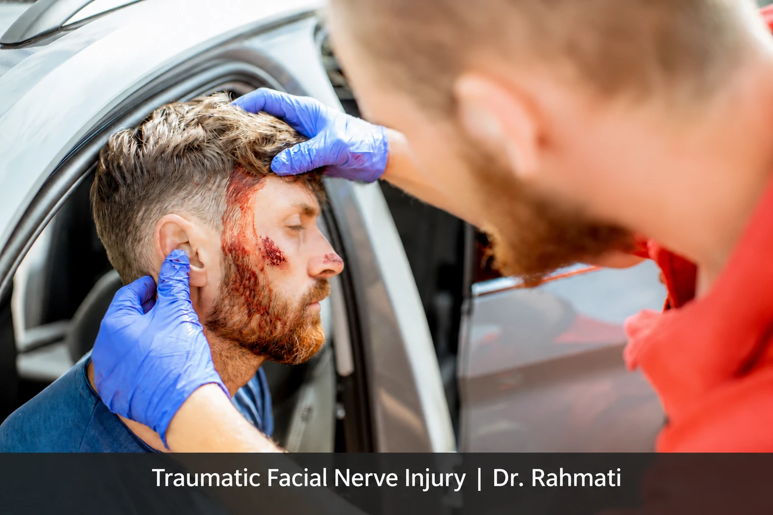
Traumatic Facial Nerve Injury can follow a road collision, a fall, a blow to the head, or a deep facial wound. Some injuries temporarily interrupt nerve signals; others damage the internal fibers or divide the nerve. A drooping mouth or an eye that will not close therefore needs assessment rather than an assumption about recovery or surgery. The appearance of the face alone cannot establish whether a nerve has been cut.
Care begins with potentially dangerous injuries to the head, neck, breathing and circulation. Facial movement and eye protection are then addressed alongside other problems. This article explores one of the conditions discussed in our guide to facial paralysis causes. It explains the assessment process, but cannot determine which medicine or operation an individual needs without examination.
How does Traumatic Facial Nerve Injury happen?
The facial nerve carries movement signals to the forehead, eyelids, cheeks and lips. Part of its course passes through the temporal bone beside structures of the ear. Head trauma can produce a fracture, compression, stretching or swelling around this pathway. Outside the skull, a deep wound near the ear or across the cheek can damage one or several branches.
In a less severe injury, the nerve remains connected but conduction is disrupted. Other injuries damage the internal fibers, and some divide the nerve completely. These patterns have different treatment implications. The clinician combines the circumstances of the accident with examination, imaging and selected electrical tests. Neither a dramatic facial droop nor a single test result reliably describes every aspect of the injury.

When emergency care comes first
Reduced consciousness, a seizure, repeated vomiting, worsening headache, limb weakness, speech difficulty, breathing problems or uncontrolled bleeding after an accident require urgent assessment. Neck pain and possible spinal injury also matter. Do not wait for a routine facial clinic appointment while these symptoms are present. Avoid unnecessary movement of someone with a possible neck injury and follow emergency service instructions.
Blood or clear fluid coming from the ear, new hearing loss and severe dizziness should also be reported promptly. They do not establish a diagnosis by themselves, but can accompany temporal bone or neighboring injuries. Do not insert objects into the ear or forcefully plug drainage. The emergency team first stabilizes the patient, then arranges the appropriate assessment of the ear, hearing and facial nerve.
Immediate weakness versus delayed weakness
Weakness present immediately after the impact raises concern about a significant injury, but does not prove nerve division. In other patients, weakness develops over subsequent hours or days as swelling or inflammation evolves. Clinicians need to know when it was first noticed, whether movement was completely absent, and whether function is improving or deteriorating.
The onset may be uncertain when a person was unconscious or receiving sedating medicines. Relatives should describe what they actually observed rather than guess a precise time. Earlier photographs or videos can help establish baseline movement, but collecting them must not delay care. The examination also checks whether brain injury or other nerve problems contribute to the facial change.
Symptoms and the first examination
Possible findings include reduced eyebrow movement, incomplete eye closure, a lowered mouth corner, leakage of drinks and an altered smile. A branch injury may affect one region while movement elsewhere is preserved. Our article on facial nerve palsy symptoms describes these functional changes. Their interpretation after trauma depends on the mechanism and associated injuries.
The clinician examines movement at rest and during activity, eye closure, wounds, the ear, hearing and associated neurological signs. Photographs and a documented facial grading system help compare later visits. A home forehead test cannot reliably locate the injury or exclude a brain problem. Repeat examinations can reveal changes that a brief assessment immediately after the accident cannot capture.
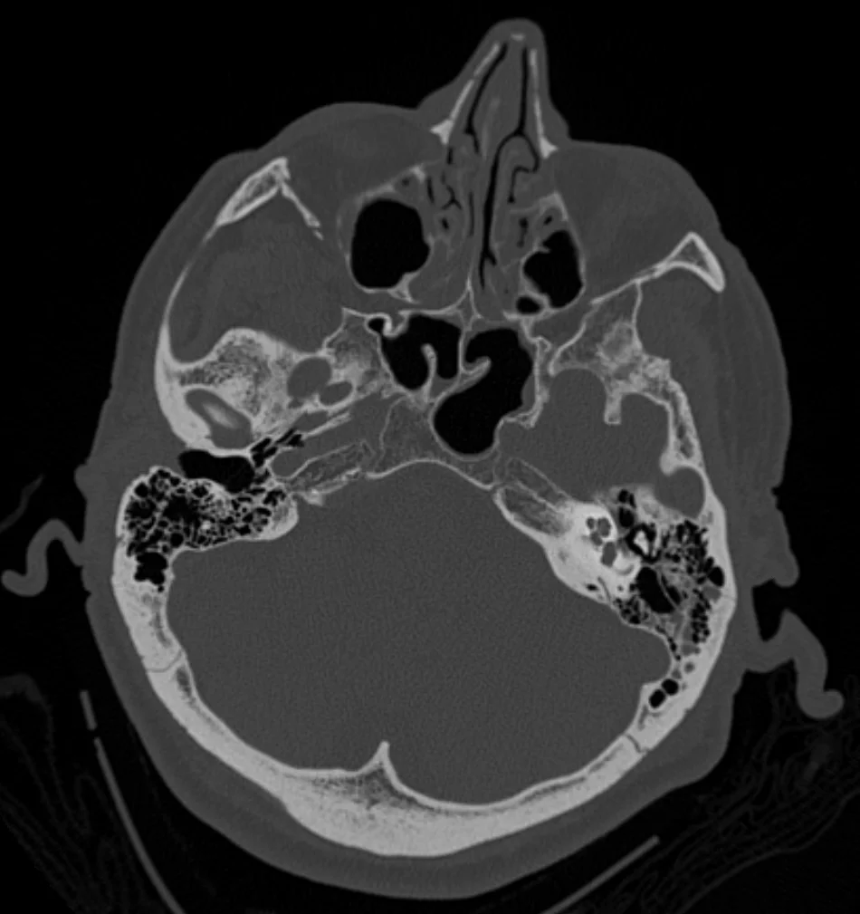
Clinical CT image published as a temporal bone fracture; this scan alone does not establish facial nerve damage. Credit: James Heilman, MD. Image source; CC BY-SA 4.0. Converted to WebP with proportions preserved; the image retains the same license.
What imaging and electrical tests can tell us
Head CT helps assess selected acute injuries, while high resolution temporal bone CT can show fracture details. Not everyone needs the same imaging package. The choice depends on ear symptoms, the degree of weakness, existing scans and the question that could change treatment. MRI may be useful in particular circumstances for soft tissues or alternative diagnoses. Imaging does not directly measure every element of nerve function.
Electroneuronography and electromyography can provide information about nerve responses and muscle activity. Timing matters: a very early test may not represent the eventual extent of damage. A percentage on a report should not automatically become a surgical instruction without the examination, onset history and fracture findings. Selected repeat testing can clarify progress when it answers a specific clinical question.

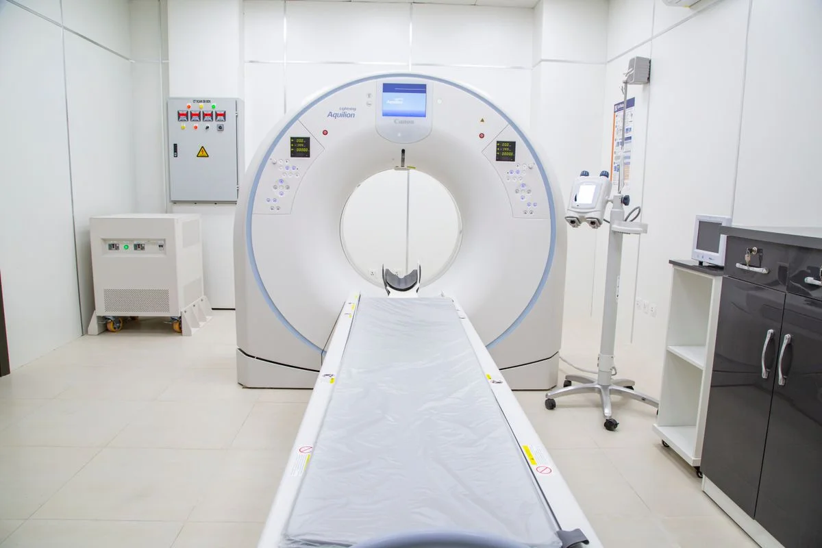
Real CT scanner shown to illustrate diagnostic imaging, not a patient or equipment belonging to Dr. Rahmati. Source and photographer profile on Pexels: contributor displayed as contact me +923323219715. Pexels License. Converted to WebP without changing proportions.
Does every patient need surgery?
No. When nerve continuity is preserved, eye protection, treatment chosen by the clinician and scheduled monitoring may be appropriate. A prospective study by Thakar and colleagues reported recovery with nonsurgical care in a selected group with undisplaced temporal bone fractures. It was not a randomized comparison of surgery against observation, and its findings cannot be generalized to penetrating wounds or a divided nerve.
A wound suggesting nerve division, particular evidence of severe compression, or an unfavorable clinical course may require surgical evaluation. Direct repair, nerve grafting and decompression are different procedures with different purposes. The choice depends on injury location, nerve continuity, hearing and overall health. The specialist should explain the reason for a proposed operation, its alternatives and its risks, without promising complete recovery.
Early referral matters, particularly when division is suspected. However, passing a single time threshold does not mean that every reconstructive option has disappeared. Much of the surgical evidence comes from small or retrospective groups with differing injuries. Decisions therefore require individual judgment. If monitoring is chosen, it should include documented findings, a review date and clear reasons to change the plan.
Protecting the eye and managing daily activities
An eyelid that does not close can leave the cornea dry and vulnerable even before severe discomfort appears. The care team advises on lubrication, overnight protection and whether an eye specialist or eyelid procedure is needed. Eye pain, marked redness, light sensitivity or reduced vision need prompt assessment. Do not postpone protection until the smile improves, and learn the correct technique rather than assume that any eye patch is sufficient.
Weak lip control can affect drinking, eating and mouth care. Discuss practical adjustments with the team, and report new choking or swallowing problems rather than automatically attributing them to the face. Sleep, work and social confidence may also be affected. Addressing these concerns is part of treatment; it does not imply that the effects are purely psychological or that appearance is unimportant.
Rehabilitation and realistic expectations
Rehabilitation should match the stage of recovery and the movement pattern. Forceful exercises, vigorous massage and unsupervised electrical stimulation are not substitutes for specialist assessment. Unwanted linked movements, such as eye narrowing during a smile, may represent synkinesis and need a tailored approach. Useful training emphasizes control, coordination and comfort rather than producing the strongest possible contraction regardless of its pattern.
Recovery depends on the type of injury, the distance to the muscles, general health and examination findings. No visible change within one week does not by itself establish treatment failure. Equally, indefinite waiting without review is inappropriate. Agree on follow-up arrangements and warning signs. Persistent weakness may lead to consideration of nerve reconstruction, muscle-based reanimation or tissue support, according to the remaining function.
Frequently asked questions
Does every temporal bone fracture cause facial paralysis?
No. Fracture location and its relationship to the nerve matter. The presence of a fracture alone cannot predict the degree of facial movement loss.

Should I start steroids myself?
No. Medication depends on the injury, associated conditions and contraindications. Steroids cannot reconnect a divided nerve and are not suitable for every accident patient.
Is assessment still worthwhile after a delay?
Yes. Earlier evaluation is preferable, but repair or reanimation options depend on the nerve and muscles. A delay does not automatically make all treatment futile.
What should I bring to the appointment?
Bring the accident records, scan files and reports, medication list, an account of when weakness appeared, and details of changes in hearing, balance or vision.
Conclusion
Facial weakness after an accident ranges from a temporary conduction problem to a serious nerve injury. Stabilizing associated injuries, protecting the eye, identifying the damage and tracking function are the priorities. Treatment should follow the evidence for the individual patient rather than a photograph, a single number or a guaranteed recovery timetable.


