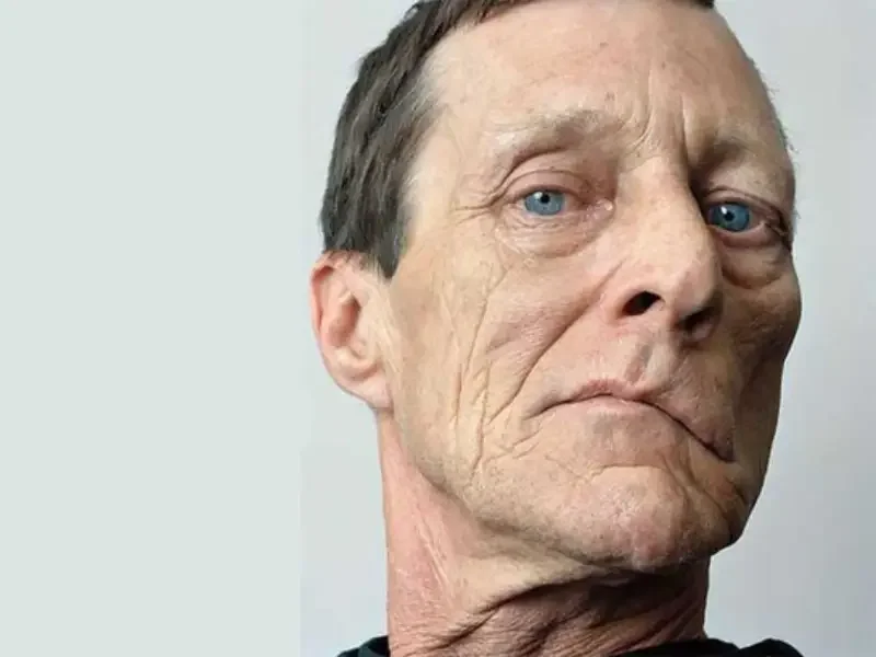
What is gracilis transfer for smile reanimation?
Smile Reanimation With Gracilis Muscle Transfer is a dynamic reconstructive operation for selected people who have lost meaningful movement at the corner of the mouth after facial paralysis. The surgeon transfers a segment of gracilis muscle from the inner thigh together with its supplying vessels and motor nerve. Using microsurgical techniques, the muscle is attached in the face, its blood supply is restored, and its nerve is connected to a functioning motor input. The aim is to create a new source of active pull at the corner of the mouth, not to promise restoration of a previously normal smile.
A smile involves complex, coordinated movements of several facial muscles. Even after a technically successful transfer, resting symmetry, speech, lip control, emotional expression, and the spontaneity of smiling may improve to different degrees. The assessment begins by asking which limitations matter most to the patient. Standardized photographs, recorded movements, a history of previous treatment, and careful examination establish realistic goals rather than assuming that one procedure can address every consequence of facial paralysis.
Why longstanding paralysis can require a new muscle
After injury to the facial nerve, denervated facial muscles can weaken, shrink, and progressively lose their ability to respond to new nerve signals. Early in the course of paralysis, direct nerve repair, nerve grafting, or nerve transfer may be appropriate in selected circumstances because native muscles may still be viable. If the original smile muscles have undergone profound chronic changes, restoring nerve traffic alone may not generate useful motion. Transferring a living, innervatable muscle offers a different route to dynamic reconstruction.
The cause and duration of paralysis are crucial but never tell the whole story. Tumor surgery, trauma, congenital syndromes, and some neurological disorders may produce very different patterns of weakness. Patients with recent facial palsy should not assume that free muscle transfer is their next step; spontaneous recovery and less invasive alternatives may need assessment. A careful distinction between movement that is absent, weak, or abnormally coordinated prevents choosing an operation that is more extensive than necessary.
Understanding the gracilis donor muscle
The gracilis is a long, relatively slender muscle along the inner thigh that helps adduct the hip and contributes to knee function. Other muscles share many of these tasks, which makes carefully planned gracilis harvest possible in reconstructive surgery. Nevertheless, removing it is not completely without consequences. Patients may experience thigh discomfort, swelling, scars, altered sensation, and temporary functional limitations, particularly during early recovery or sporting activity.
For reconstruction, the muscle has useful length, a defined blood supply, and a motor nerve that can be connected to a facial donor nerve. The surgeon chooses how much muscle to harvest and where to attach it according to facial anatomy and the desired direction of pull. An anatomical image of the thigh is not a photograph of an accomplished smile reconstruction; clinical images must distinguish the site of muscle harvest from the actual facial outcome.

Who might be a candidate?
Free functional muscle transfer is commonly considered in established paralysis when the original smile muscles are unlikely to recover enough function. Suitability also depends on blood vessels, the availability of a motor nerve, prior surgery or radiation, medical conditions, smoking history, and whether a lengthy microsurgical operation is safe. Age alone cannot decide candidacy. Patients with bilateral paralysis, congenital facial weakness, or major facial scarring require additional individual planning.
The patient and surgeon should agree on the principal goal: movement at the mouth corner, improved symmetry, greater ease in social expression, or a combination of these. Reconstructing a smile does not automatically solve inability to close an eye or weakness of the forehead. A balanced discussion covers nonsurgical care, static support procedures, nerve-based strategies, and muscle transfer, along with possible staged treatment. The site’s discussion of muscle transfer in smile reanimation offers additional context.
Preoperative assessment and planning
At consultation, the team examines facial movement at rest and with smiling, lip closure, cheek tone, prior scars, and the movement of the healthy side. Short videos while speaking, smiling gently, and showing the teeth can capture information that still photographs miss. Selected patients may need electrophysiological testing or imaging based on the cause and timing of paralysis. No single test or fixed diagnostic pathway is appropriate for everyone.
Surgical planning includes selecting a donor nerve, suitable facial vessels, attachment points, and a recovery strategy. Previous parotid surgery, neck dissection, radiation treatment, and existing nerve grafts may change the available options. It is particularly important to discuss what a voluntary bite-activated smile might feel like compared with an emotionally triggered smile. Setting expectations before the operation can help patients evaluate meaningful improvement without requiring impossible perfection.
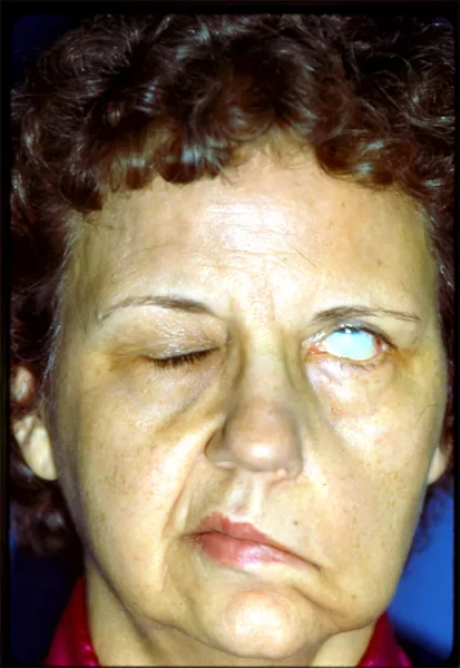
Educational photograph of facial paralysis following acoustic neuroma surgery; not a gracilis transfer result or a patient of Dr Rahmati. Student30 — Wikimedia Commons; CC BY-SA 4.0. Converted to WebP; resized.
How the operation is performed
The operation involves harvesting a tailored gracilis muscle segment with its vascular pedicle and motor nerve from the inner thigh. Through carefully planned facial incisions, the surgeon places the muscle and secures it so its eventual contraction draws the corner of the mouth in an appropriate direction. Under magnification, blood vessels are joined to recipient vessels in the face or neck. Restoring reliable circulation is critical because the transplanted muscle must remain alive for any later nerve-driven movement to occur.
The gracilis motor nerve is then joined to the selected donor nerve. After checking circulation and positioning, wounds are closed and a postoperative monitoring plan begins. The exact approach varies with facial anatomy, previous treatment, and whether the procedure is part of a staged reconstruction. The operation creates the possibility of movement; it does not provide a new smile immediately. Functional recovery depends on nerve regeneration, muscle adaptation, and sustained rehabilitation.
Choosing the masseteric nerve or a cross-face graft
The masseteric nerve, which normally activates a chewing muscle, provides a relatively strong motor source for the transferred gracilis. Patients may initially learn to produce movement by gently activating the jaw. With rehabilitation, the degree to which that movement becomes useful during ordinary smiling can increase. The masseteric route often allows one-stage transfer, although it does not guarantee a spontaneous emotional smile.
A cross-facial nerve graft carries signals from branches of the functioning facial nerve on the opposite side. This may offer a pathway for more natural coordination, but nerve growth takes time and a staged operation is often required. Some teams use dual innervation to combine potential advantages. A systematic review of published studies found differences in measured movement between donor strategies but insufficient consistent reporting to declare one option definitively best for all patients. The related article on masseteric nerve transfer versus cross-face grafting explores the decision.
One-stage and two-stage reconstruction
In a common one-stage strategy, the gracilis is transferred and connected to the masseteric nerve during the same procedure. When cross-facial nerve grafting is planned, the surgeon may first place a nerve graft from the intact side and allow time for nerve fibers to grow across it. A later operation transfers and connects the gracilis. The staged pathway therefore includes more than one healing period and a longer interval before potential new movement becomes visible.
Faster is not synonymous with better for every person. Donor nerve quality, age, other illnesses, the function of the unaffected side, the history of prior facial surgery, and preferences about voluntary versus emotional activation all matter. Patients should ask how many operations are anticipated and what events might cause the plan to change. Even when an initial nerve graft appears successful, definitive muscle performance cannot be guaranteed in advance.
Immediate recovery and flap monitoring
The first postoperative days focus on maintaining reliable blood supply to the transferred muscle and healing the facial and thigh wounds. Clinicians watch for problems with circulation, bleeding, and infection, while also addressing swelling and pain. Instructions about activity, wound care, medication, and when to contact the team should be individualized. Fever, escalating pain, unexpected bleeding, or other concerning symptoms warrant prompt clinical advice rather than self-treatment.

Early swelling or a changed resting contour is not a measure of the final functional outcome. The new muscle generally cannot contract meaningfully before nerve fibers reach it and functional connections develop. Published clinical series describe the first movements after a period of months, but the timing varies substantially. Scheduled visits help identify complications and guide decisions about starting or adjusting rehabilitation.
Rehabilitation and motor learning
Once the surgical team confirms that activation is appropriate, a therapist can help the patient learn to recruit the transferred muscle. For masseteric innervation, early exercises sometimes pair jaw activation with an attempted smile. Visual feedback, targeted movements, and careful control of unwanted compensations may help shape a useful facial expression. Exercise should start at a clinician-approved stage because excessive or poorly timed movement may cause discomfort or undermine recovery instructions.
Rehabilitation is more than maximizing millimeters of mouth-corner excursion. It may address symmetry, speech-related movement, confidence during conversations, and the ability to use the new smile without concentrating on each motion. Progress can be tracked with standardized recordings under similar conditions. People differ considerably in the extent to which a deliberate movement becomes integrated into everyday emotional expression. Long-term follow-up remains valuable after the first detectable contraction.
Expected benefits and uncertainty in outcomes
Studies of free gracilis transfer suggest that selected patients can gain mouth-corner movement, better symmetry during smiling, and improvement in some patient-reported quality-of-life measures. Yet research groups differ in diagnosis, age, donor nerve, duration of paralysis, and methods of measuring outcomes. An average improvement reported by a center should never be treated as a guaranteed personal result.
A systematic review and meta-analysis in JAMA Otolaryngology–Head & Neck Surgery examined donor nerve options and found evidence suggesting greater measured excursion with masseteric innervation in some comparisons, while highlighting major limitations from inconsistent outcome reporting. Other research has examined potential advantages in emotional spontaneity for cross-facial and dual-innervation approaches. A useful outcome discussion considers resting position, movement amplitude, coordination, social function, and the patient’s own priorities. Symmetrical or fully spontaneous smiling is not assured.
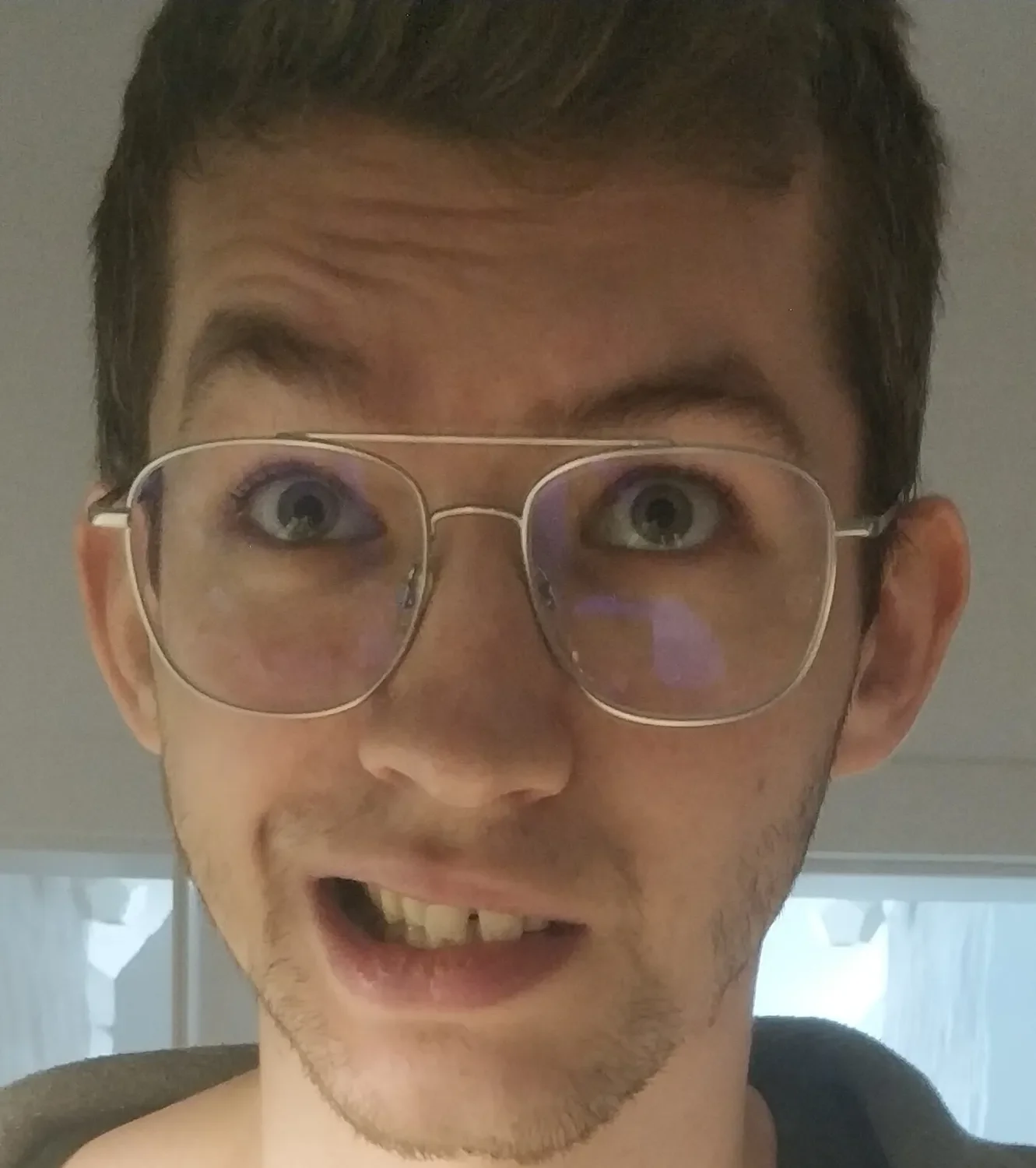
Educational photograph of Bell’s palsy and facial asymmetry; not a gracilis transfer result or a patient of Dr Rahmati. Wdobrzanski — Wikimedia Commons; CC BY-SA 4.0. Converted to WebP; resized.
Surgical risks and donor-site effects
Free functional muscle transfer is a significant microsurgical operation. Potential complications include infection, hematoma, vascular thrombosis, compromised flap blood supply, failure of the transplant, nerve-related problems, asymmetry, unwanted bulk, scars, and the possible need for revision surgery. General anesthetic risk and the burden of a longer operation must also be considered. The likelihood and management of complications depend on individual circumstances and the care setting.
At the thigh, patients may experience discomfort, swelling, scarring, numbness, and temporary weakness. Most people can adapt to the loss of a carefully harvested gracilis, but outcomes vary with preexisting mobility and the extent of surgery. Before consent, patients should discuss the likelihood of additional interventions, signs requiring urgent assessment, smoking cessation, and how their broader medical conditions affect wound healing.
Dynamic muscle transfer compared with static procedures
Static techniques support or reposition tissues to improve appearance at rest but do not create an actively contracting new smile muscle. Free gracilis transfer is a dynamic technique because its purpose is to generate movement through reinnervation. Some people benefit from a combination: static suspension may address resting droop while dynamic reconstruction aims to create motion. A nerve-only procedure may be preferable when native facial muscles remain capable of responding.
Eye protection is a separate high-priority issue in facial paralysis. Difficulty closing the eyelid and corneal exposure require appropriate assessment regardless of the plan for the lower face. Smile surgery should not postpone necessary eye care. Understanding these distinct goals reduces the risk of expecting a single operation to fix every region of the face. The site’s overview of facial nerve transfer discusses alternative nerve-based strategies.
Common patient questions
Will removing gracilis stop me from walking normally? Other thigh muscles usually compensate for much of its function, but temporary weakness, pain, or activity restrictions can occur. Will there be visible scars? Both the thigh harvest and facial access leave scars, whose appearance varies. Does the operation create movement straight away? No; nerve growth and motor relearning take time. Can surgery help after years of paralysis? It may be considered in longstanding cases, subject to health, anatomy, and nerve availability.

Will the new smile be spontaneous? This cannot be promised; the nerve source and individual rehabilitation response influence it. Can I avoid follow-up therapy? Rehabilitation is usually an important component of treatment and should be discussed before consenting to surgery. Is the operation purely cosmetic? Its goals often include functional movement and communication, not only facial appearance. Individual questions should be answered in an appointment rather than by relying on generalized online outcome images.
Conclusion: individualized smile reanimation
A free gracilis transfer can offer a new source of dynamic movement to the corner of the mouth in selected people with established facial paralysis. Its value depends on choosing the right candidate, selecting a suitable donor nerve, positioning the muscle correctly, restoring its circulation, and supporting recovery through rehabilitation. It is not the same as simply repairing the facial nerve and it is not the right choice for every case of weakness.
A specialist consultation should review the diagnosis, duration of paralysis, previous operations, eye protection, alternative treatments, expected timeline, and both surgical and donor-site risks. Informed consent includes acknowledging that improvement is possible without promising a perfectly symmetrical or spontaneous smile. This article is educational and does not substitute for examination and individualized medical advice.
Medical references
https://pmc.ncbi.nlm.nih.gov/articles/PMC7099528/