
Facial Nerve Injury After Surgery may cause difficulty raising an eyebrow, closing an eye or moving the corner of the mouth. New weakness does not automatically mean that the nerve was cut or that paralysis will be permanent. Some disturbances are temporary; others require prompt investigation for a more substantial injury. The first priority is to contact the surgical team and describe exactly when movement changed.
Postoperative weakness is one possible cause of facial dysfunction, not the only explanation. The parent article on diseases causing facial paralysis discusses other conditions. This guide helps patients prepare for assessment and understand care options. It cannot decide whether an individual needs another operation or provide a medication plan without examination and review of the original surgery.
How can Facial Nerve Injury After Surgery occur?
The facial nerve supplies many muscles of expression and travels near the brainstem, through the temporal bone and into the parotid region. During nearby surgery, stretching, pressure, heat or changes in blood supply may disturb its function. The consequences depend on the mechanism, location and extent of injury rather than simply on the fact that an operation took place.
An anatomically continuous nerve with impaired conduction is different from a nerve that has been divided. In some cancer operations, removing an involved nerve segment is part of planned disease treatment. Weakness alone therefore cannot establish what happened or prove a surgical error. The operative findings and current examination must be considered together before choosing treatment.

Which operations can be associated with weakness?
Relevant procedures include parotid operations, some middle-ear and mastoid surgery, skull-base tumor removal and selected facial procedures. Weakness may involve an entire side or only certain branches. Reduced lower-lip movement and incomplete eye closure are different functional problems, so their assessment and protective measures cannot simply be treated as identical.
The original disease, previous operations and preoperative nerve function influence interpretation. Removing a growth involving the nerve is not comparable to a limited operation away from it. Our guide to a facial nerve tumor explains this relationship. A complication rate from one procedure should not be applied to every operation or used to predict an individual outcome.
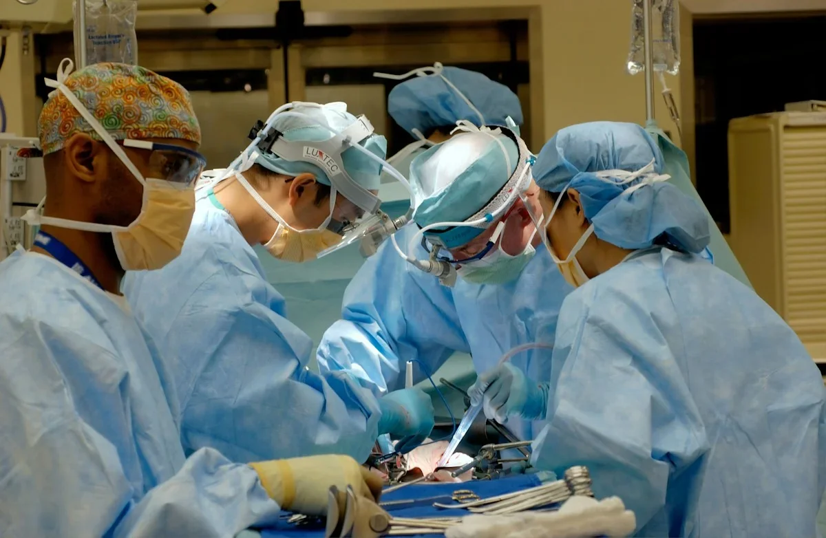
Real general operating-room photograph; it does not identify facial nerve surgery or demonstrate a nerve injury. The people shown are not Dr. Rahmati’s team. National Cancer Institute / Unsplash, Unsplash License; resized and converted to WebP.
Immediate versus delayed facial weakness
Weakness noticed immediately after an operation needs comparison with the preoperative examination and operative record. Local anesthetic effects can be considered in certain settings, but patients should not assume that medication explains every new change. Complete weakness or concern about nerve division calls for prompt specialist review rather than waiting for a routine follow-up appointment.
Sometimes movement is initially satisfactory and deteriorates days later. Inflammation or other mechanisms may be considered, but “delayed” does not mean harmless. The team must assess the wound and consider relevant problems such as a hematoma or infection. Record the last time movement appeared normal and whether the weakness has remained stable, fluctuated or progressed.
When should you seek help urgently?
Report new or worsening postoperative facial weakness to the surgeon or treating center the same day. Explain whether the eyelid closes and whether drinking or mouth control has changed. Rapid swelling, bleeding, breathing difficulty or swallowing difficulty requires urgent assessment. Do not wait for an ordinary appointment when symptoms suggest a potentially serious postoperative problem.
Sudden facial drooping with arm or leg weakness, abnormal speech, visual change or imbalance requires immediate contact with the local emergency service. Surgery does not exclude another neurological emergency. Eye pain, marked redness, light sensitivity or reduced vision also needs prompt attention. Protecting sight should not be postponed until facial movement improves.
What do examination and tests show?
Assessment examines forehead movement, eyelid closure, smiling and lip control. Preoperative photographs or videos and the earliest postoperative examination can provide useful comparisons. House–Brackmann and Sunnybrook scores help track movement, but everyday concerns such as leaking drinks, speech difficulty or eye discomfort also need to be recorded. A single score cannot describe all the patient’s needs.
The operation report may describe whether the nerve remained continuous, responded to stimulation or underwent reconstruction. Monitoring findings provide useful context but do not guarantee that postoperative weakness will never develop. Reviewing the record is intended to connect the surgical observations with the current pattern of dysfunction, not to replace clinical examination.
Electrical nerve and muscle studies may help assess severity and signs of recovery when performed at an appropriate time. A very early result may not show the eventual extent of injury, and serial assessment can sometimes be needed. Imaging is selected for a specific question; high-resolution temporal-bone CT may be useful after some middle-ear surgical injuries.

Eye protection starts immediately
An eyelid that does not close fully exposes the cornea to drying and injury. The clinical team can advise suitable lubrication and teach safe nighttime protection. Taping or using a protective device requires the correct technique. Simply covering the eye does not guarantee that the eyelid is closed, and inappropriate pressure or application can create further problems.
A watering eye is not necessarily a protected eye. Discomfort or visual changes should prompt review, and an ophthalmic assessment may be necessary. Drops, ointment or devices recommended for another person may not be suitable for you. Ask for clear instructions, including what to do if symptoms worsen between appointments.
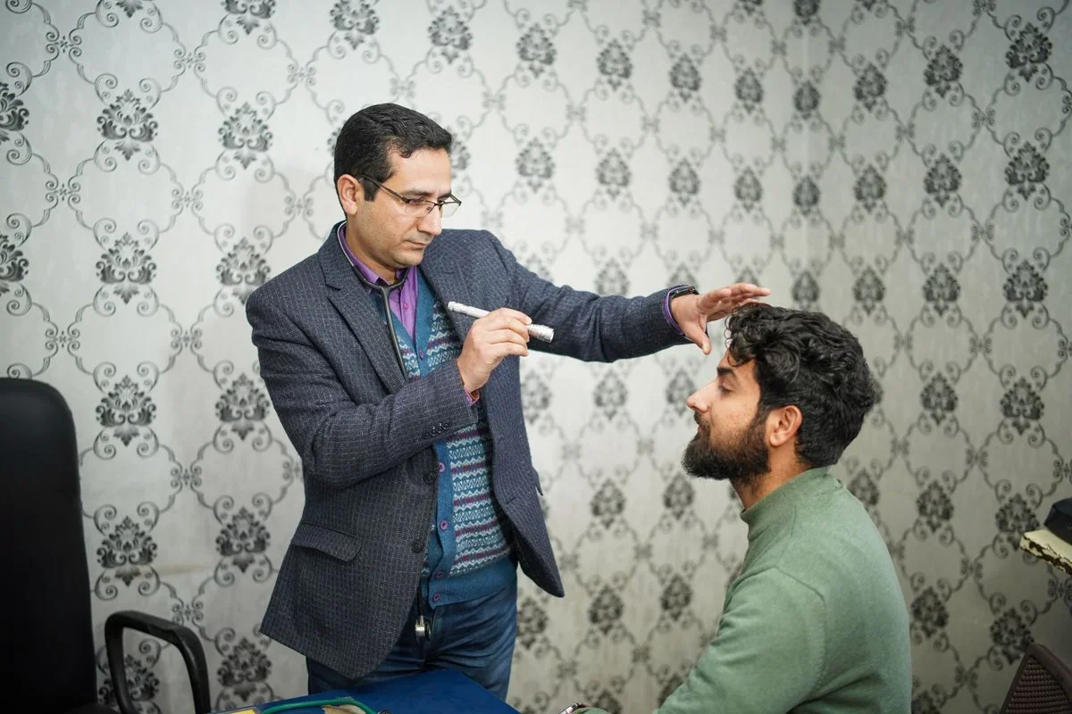
Real stock photograph illustrating an eye examination, not a person diagnosed with facial nerve injury or a patient of Dr. Rahmati. Imad Clicks / Pexels, Pexels License; resized and converted to WebP.
Observation or nerve repair?
Active observation can be appropriate when continuity is preserved and the assessment suggests a potentially temporary disturbance. This decision should follow evaluation. Observation includes a planned examination date, documentation of movement and clear reasons to reconsider treatment. A general instruction to wait several months without assessment or monitoring is not an adequate individualized plan.
Known or suspected nerve division warrants early referral to a team experienced in facial nerve repair. Direct tension-free repair or an interposition graft may be considered in suitable injuries. Timing depends on the operative situation and tissue condition. A later presentation does not mean that all options have disappeared; seek specialist assessment rather than assuming an arbitrary deadline has ended care.
Steroids or antiviral medicines are not a shared treatment for every postoperative weakness, and they cannot reconnect a physically divided nerve. Prescribing depends on the likely cause and the patient’s circumstances. Do not start treatment from another patient’s prescription or change existing medication without discussing it with the treating team.
Rehabilitation and realistic recovery goals
Specialist facial retraining may help selected patients control movement, but the program should fit the stage of recovery and wound restrictions. Forceful exercise, painful massage or unsupervised electrical stimulation is not a guaranteed shortcut. If the eye narrows while smiling or movements become linked, assessment for synkinesis may be appropriate rather than simply increasing exercise intensity.
Persistent weakness may lead to discussion of nerve transfer, muscle transfer or supportive procedures according to remaining muscle function. Eye closure, oral control and symmetry at rest are separate goals. Improvement may be gradual and incomplete. Another patient’s result or a small study cannot guarantee your outcome, and emotional concerns about appearance and social contact deserve attention too.
Frequently asked questions
Is postoperative facial weakness always permanent?
No. Some disturbances are temporary. Appearance alone cannot establish injury severity; the mechanism and progression need assessment.

Does an intact nerve in the operative record exclude weakness?
No. Anatomical continuity and immediately normal function are different findings. Examination and follow-up remain important.
Does every patient need another operation?
No. The cause, location and severity guide decisions. Suspected nerve division requires prompt specialist evaluation.
What should I bring to the appointment?
Bring the operation report, relevant pathology, earlier images, medication details and a timeline of changing symptoms.
Conclusion
Care for Facial Nerve Injury After Surgery begins with prompt reporting, eye protection and review of the operation. Observation, repair and reconstruction should be selected according to the injury and the patient’s functional goals, with a clear follow-up plan.
Medical references
- Acute nerve repair, 2022
- Middle-ear surgical injury, 2023
- Post-facelift paralysis, 2024
- Delayed postoperative paralysis, 2019
- Delayed palsy after schwannoma surgery, 2024
- Molinari et al., rehabilitation, 2024
- Facial exercise randomized trial, 2016
- Nerve graft rehabilitation, 2025
- Prospective nerve monitoring study, 2022
- NHS: eye protection
- NHS: urgent eye symptoms
- CDC: neurological emergency signs


